Showing posts with label Microscopes. Show all posts

Thursday, March 19, 2009

Beam's Path through the Column in Scanning Electron Microscope (SEM)

Beam's Path through the Column in Scanning Electron Microscope (SEM)
A beam of electrons is generated in the electron gun, located at the top of the column, which is pictured to the left. This beam is attracted through the anode, condensed by a condenser lens, and focused as a very fine point on the sample by the objective lens. The scan coils are energized (by varying the voltage produced by the scan generator) and create a magnetic field which deflects the beam back and forth in a controlled pattern. The varying voltage is also applied to the coils around the neck of the Cathode-ray tube (CRT) which produces a pattern of light deflected back and forth on the surface of the CRT. The pattern of deflection of the electron beam is the same as the pattern of deflection of the spot of light on the CRT.

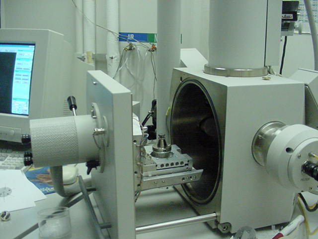
The electron beam hits the sample, producing secondary electrons from the sample. These electrons are collected by a secondary detector or a backscatter detector, converted to a voltage, and amplified. The amplified voltage is applied to the grid of the CRT and causes the intensity of the spot of light to change. The image consists of thousands of spots of varying intensity on the face of a CRT that correspond to the topography of the sample.

The Electron Source of Scanning Electron Microscope (SEM)

The Electron Source of Scanning Electron Microscope (SEM)
The electron beam comes from a filament, made of various types of materials. The most common is the Tungsten hairpin gun. This filament is a loop of tungsten which functions as the cathode. A voltage is applied to the loop, causing it to heat up. The anode, which is positive with respect to the filament, forms powerful attractive forces for electrons. This causes electrons to accelerate toward the anode. Some accelerate right by the anode and on down the column, to the sample. Other examples of filaments are Lanthanum Hexaboride filaments and field emission guns.

Scanning Electron Microscope (SEM) Process and image formation

File:SEM chamber1.JPG
In a typical SEM, an electron beam is thermionically emitted from an electron gun fitted with a tungsten filament cathode. Tungsten is normally used in thermionic electron guns because it has the highest melting point and lowest vapour pressure of all metals, thereby allowing it to be heated for electron emission, and because of its low cost. Other types of electron emitters include lanthanum hexaboride (LaB6) cathodes, which can be used in a standard tungsten filament SEM if the vacuum system is upgraded and field emission guns (FEG), which may be of the cold-cathode type using tungsten single crystal emitters or the thermally-assisted Schottky type, using emitters of zirconium oxide.

The electron beam, which typically has an energy ranging from a few hundred eV to 40 keV, is focused by one or two condenser lenses to a spot about 0.4 nm to 5 nm in diameter. The beam passes through pairs of scanning coils or pairs of deflector plates in the electron column, typically in the final lens, which deflect the beam in the x and y axes so that it scans in a raster fashion over a rectangular area of the sample surface.

When the primary electron beam interacts with the sample, the electrons lose energy by repeated random scattering and absorption within a teardrop-shaped volume of the specimen known as the interaction volume, which extends from less than 100 nm to around 5 µm into the surface. The size of the interaction volume depends on the electron's landing energy, the atomic number of the specimen and the specimen's density. The energy exchange between the electron beam and the sample results in the reflection of high-energy electrons by elastic scattering, emission of secondary electrons by inelastic scattering and the emission of electromagnetic radiation, each of which can be detected by specialized detectors. The beam current absorbed by the specimen can also be detected and used to create images of the distribution of specimen current. Electronic amplifiers of various types are used to amplify the signals which are displayed as variations in brightness on a cathode ray tube. The raster scanning of the CRT display is synchronised with that of the beam on the specimen in the microscope, and the resulting image is therefore a distribution map of the intensity of the signal being emitted from the scanned area of the specimen. The image may be captured by photography from a high resolution cathode ray tube, but in modern machines is digitally captured and displayed on a computer monitor and saved to a computer's hard disc.

Scanning Electron Microscope (SEM)

Scanning Electron Microscope (SEM)
The Scanning Electron Microscope (SEM) is a microscope that uses electrons rather than light to form an image. There are many advantages to using the SEM instead of a light microscope.

The scanning electron microscope (SEM) is a type of electron microscope that images the sample surface by scanning it with a high-energy beam of electrons in a raster scan pattern. The electrons interact with the atoms that make up the sample producing signals that contain information about the sample's surface topography, composition and other properties such as electrical conductivity.

The SEM has a large depth of field, which allows a large amount of the sample to be in focus at one time. The SEM also produces images of high resolution, which means that closely spaced features can be examined at a high magnification. Preparation of the samples is relatively easy since most SEMs ony require the sample to be conductive. The combination of higher magnification, larger depth of focus, greater resolution, and ease of sample observation makes the SEM one of the most heavily used instruments in research areas today.

Friday, February 13, 2009

Spencer in profile


A Spencer microscope, by American Optical.
Not sure of the precise vintage, but somewhere circa 1950's.

Tuesday, January 6, 2009

Microscope Celestron Professional Biological by Celestron

Microscope Celestron Professional Biological by Celestron
Celestron optical instruments deliver excellent resolution and contrast. Celestron's instruments offer versatility in viewing any microscopy subject that interests you. Celestron offers microscopes suited for a wide variety of uses - hobby, education and research. This is a heavy duty professional level biological microscope. An extra large mechanical stage allows ample working space and knobs control both axes for easy centering of specimen slides. Two sets of wide angle eyepieces allow eight different powers. All metal construction of the bases, arms, stands, and heads give the microscope lasting durability and rigidity. It has five prepared slides included so you can begin enjoying viewing right "out from the box". The Microscope is backed with a two year limited warranty.

Technical Details

* A large, heavy duty professional level microscope that provides powers up to 1500x for very serious users for analysis and research
* 10x & 15x wide field (18mm & 13mm fields of view) eyepieces and 4x, 10x, 40x, & 100x objective lenses combine give powers of 40x, 60x, 100x, 150x, 400x, 600x, 1000x &1500x
* Binocular head rotates 360° which allows comfortable two eye viewing at any position plus enables others to share your view without moving the microscope
* 45° inclined viewing angle provides comfortable viewing
* Built-in halogen illumination provides direct control of light with brightness adjustment
See more detail

Microscope Meade 9600 by Meade

Microscope Meade 9600 by Meade
Model 9600–Includes 360 degrees -rotatable binocular head with interpupillary distance adjustment; three professional-size, turret-mounted objective lenses (10x, 40x, and 100x); two wide-field eyepieces each of 10x and 12.5x for effective observing magnifications of 100x, 125x, 400x, 500x, 1000x, and 1250x; precision coarse-and-fine rack-and-pinion focusing mechanism; Abbe-type condensing lens system and iris diaphragm; electric illuminator with diffusion lens; specimen stage 110mm x 120mm with micrometric x - y controls (25mm longitudinal, 50mm transverse) for specimen centering; slide set with 5 prepared slides and 5 blank slides.

From the Manufacturer
The Meade Model 9600 biological microscope provides a virtually endless observational opportunity for the serious amateur microscopist. Similar in design and features to the Model 9460, the Model 9600 includes an important addition: a fully adjustable binocular viewing head that greatly relieves eye strain over extended observing sessions.

Examine specimens with three professional-size, turret-mounted objective lenses of 10x, 40x, and 100x magnifications; each objective lens is seated in a chromed, machined brass housing. In conjunction with eyepieces of 10x and 12.5x the Model 9600 yields effective observing magnifications of 100x, 125x, 400x, 500x, 1000x, and 1250x. Precision coarse-and-fine focusing controls permit extremely sharp focus of the image. The Model 9600's large 110mm x 120mm stage is complemented with micrometric x - y controls that permit 25mm longitudinal and 50mm transverse motions for object-centering. The 360°-rotatable binocular head allows for comfortable interpupillary adjustment; the built-in electric illuminator and diffusion lens, with Abbe-type condenser and iris diaphragm, provide bright, uniform specimen illumination.

As a microscope for the discriminating amateur scientist, or for the school or college biology laboratory, the Meade Model 9600 is designed for a lifetime of serious applications.
See more detail

Microscopes Zoom Optical

Microscopes Zoom Optical
Flexbar SMZ 1:4 Zoom Optical Body for Flexbar SMZ 1:4 Zoom Ratio Microscope. Flexbar SMZ 1:4 Ratio Stereo Zoom Microscope has an economy model high resolution Stereo Image with Zoom Magnification click stops. Featuring Greenough Zoom Optical System; 45 degree binocular stereo microscope head; wide eyepieces 10X/FN.20; magnification range 1X-4XWD=80mm; 12V/15W halogen incident illumination; 12V/10W halogen incident illumination
See more detail

Microscope Stereo Zoom by Flexbar

Microscope Stereo Zoom by Flexbar
Flexbar SMZ 1:4 Ratio Stereo Zoom Microscope. Economy model high resolution Stereo Image with Zoom Magnification click stops. Featuring Greenough Zoom Optical System; 45 degree binocular stereo microscope head; widefield eyepiece
See more detail

Microscope SMZ 1:6:7 Ratio Optical by Flexbar

Microscope SMZ 1:6:7 Ratio Optical by Flexbar
Flexbar SMZ 1:6.7 Zoom Ratio Optical Body (only) for Flexbar SMZ 1:6.7 Zoom Ratio Stereo Microscope featuring Greenough Zoom Optical System. System features 35 degree binocular stereomicroscope head, magnification range of 0.75x-5x, working distance of 113mm, a large working area stand, incident/transmitted illumination 12V/10W, and a power supply of 100V-240 (UL)
See more detail

Microscope K Series Stereo by Flexbar

Microscope K Series Stereo by Flexbar
Flexbar K Series Deluxe Stereo Microscopes. Our Highest Quality Microscopes Featuring Superb Optical Performance And Design Found On Other Brands Costing Much More. Available In Standard And Zoom Versions. Extra-Large Working Stage With Built In Incident And Transmitted Halogen Light 12V/10W With Intensity Control. 3-Way Illumination System Enhancing Choices Of Top, Bottom Or Top And Bottom Light. Main Supply 220V Ce Or 110V. Super Widefield Eyepiece 10X (Fn23) One Pair With Binocular Tube With Dioptric Com- Pensation On Both Tubes And Interpupillary Distance Adjustment Between 54mm And 76mm. Standard 1.0X Objective. Model No. 20766, Flexbar K Series Stereo Microscope Featuring 5 Step Magnification Changer At 6.4, 10, 16, 25 And 40 Ratio
See more detail

Monday, January 5, 2009

Microscopes M5 LabScope DIN by LW Scientific

Microscopes M5 LabScope DIN by LW Scientific
LW Scientific M5-P LabScope DIN Plan Lab Microscope w/ Liftime Warranty! The highly versatile M5 LabScope is built to withstand the rigors of daily laboratory use in the pathology, hematology, cytology and microbiology lab. The five-position nosepiece allows for specialized objectives, including 50X oil Plan, 60X high dry Plan, 2X Plan and Phase Contrast objectives. The oversized 10X/20 high point Plan eyepieces combined with Infinity Plan or DIN Plan objectives offer high resolution flat-field imaging over a larger specimen area. The M Series was designed for comfort and ease of operation, with all controls located ergonomically within a fingers reach. The M5 sets a new laboratory standard in microscopy. HEAD: - Binocular (Seidentopf) Head Objectives - Trinocular with C-mount or eye tube - 35mm and digital camera adapters available - Diopter adjustment - Inclined 30° (45° available), rotates 360° - 10X/20 high point eyepieces - Interpupillary distance range 50-75mm NOSEPIECE: - Reverse quintuple nosepiece - Multiple ball bearing mounted OBJECTIVE: - Infinity Plan or DIN Plan objectives, anti-fungal - 4X, 10X, 20X, 40XR, 100XR (oil) - Parfocal, parcentric, color-coded STAGE: - Mechanical stage (160mm x 140mm) - Coaxial drive controls, metal rack & pinion - Range of traverse: 77mm x 50mm - Stage locking lever - Slow-close hydraulic slide finger - Dual slide holder FOCUS: - Coarse adjustment: range of 20mm - Fine adjustment: graduation of 2µm - Tension control knob ILLUMINATION: - Moveable Abbe condenser, NA 1.25 - Iris diaphragm - Halogen light source (12V/20W bulb) - Variable light adjustment: 0-20W output - Input 90-240V / 50-60Hz automatic switching ACCESSORIES INCLUDED: - One 2 amp fuse, spare 12V/20W bulb, blue, green and yellow filters - Dust cover, immersion oil, manual and warranty card WARRANTY: - Lifetime on materials and workmanship - One year on electronic components

Technical Details

* Premium LW Scientific Lab Microscope
* 5-Posistion Nosepiece
* For laboratory use in pathology, hematology, cytology and microbiology
* Please specifiy Head when ordering.
* We carry a full line of accessories for the M5 Labscope. Please call for more information.
See more detail

Stereomicroscopes Carl Zeiss Stemi by Fisher Scientific

Stereomicroscopes Carl Zeiss Stemi by Fisher Scientific
This information applies to both the 2000/2000C models.Compact and high-performance stereomicroscope Stemi 2000. TheCarl Zeiss Stemi 2000 Stereomicroscope can be utilized for education,industrial testing, life sciences and materials research applications: Ideal for researches performed in scientific institutes and for routine quality control. Perfect for examining of large objects at low magnifications. The microscope can be used for the quality control in industry. Both models feature tapered front optics, convenient operating controls and a rounded base. 35° angle binocular tube for convenient sample viewing while providing room for specimen manipulation. Working distance: 110mm without additional objectives; 31 to 285mm with additional objectives. Built-in magnification changer for manual adjustment with a 1 to 7 continuous zoom range (see Magnification Range chart for appropriate range with each available optic and eyepiece). Front lenses can be interchanged to alter the magnification range. For photodocumentation, the Stemi 2000 includes a port for photo accessories or video camera. Both models are mounted onto a heavy metal base stand with a 11/4 in. diameter (32mm) post for optical adjustment. The Carl Zeiss Stemi Sereomicroscopes includes: Supplied with 10X/23 focusing eyepiece; additional eyepieces and auxiliary objectives available. Without illuminators; optional illuminators available; contact Superior Scientific for a quote on illumninators. Stereomicroscope Stemi 2000, 0.65X to 5.0X Zoom Range 286mm Working Distance for Stereoscopic Observation sold separately; contact Superior Scientific for more information. Technical Data: Basic instrument with changing optics. Magnification range: 6,5x - 50х / 1,95х - 225х Free working distance: 90 mm / depends on the changing optics. Field of view: 23 mm / 21 or 16 or 10 mm. Maximum object field diameter: 35m.
See more detail

Stereomicroscopes Carl Zeiss Stemi 2000C by Fisher Scientific

Stereomicroscopes Carl Zeiss Stemi 2000C by Fisher Scientific
This information applies to both the 2000/2000C models.Compact and high-performance stereomicroscope Stemi 2000c. The Carl Zeiss Stemi 2000C Stereomicroscopes can be utilized for education, industrial testing, life sciences and materials research applications: Ideal for researches performed in scientific institutes and for routine quality control. Perfect for examining of large objects at low magnifications. The microscope can be used for the quality control in industry. Both models feature tapered front optics, convenient operating controls and a rounded base. 35° angle binocular tube for convenient sample viewing while providing room for specimen manipulation. Working distance: 110mm without additional objectives; 31 to 285mm with additional objectives. Built-in magnification changer for manual adjustment with a 1 to 7 continuous zoom range (see Magnification Range chart for appropriate range with each available optic and eyepiece). Front lenses can be interchanged to alter the magnification range. For photodocumentation, the Stemi 2000C includes a port for photo accessories or video camera. Both models are mounted onto a heavy metal base stand with a 11/4 in. diameter (32mm) post for optical adjustment. The Carl Zeiss Stemi Sereomicroscopes includes: Supplied with 10X/23 focusing eyepiece; additional eyepieces and auxiliary objectives available. Without illuminators; optional illuminators available; contact Superior Scientific for a quote on illumninators. Stereomicroscope Stemi 2000, 0.65X to 5.0X Zoom Range 286mm. Working Distance for Stereoscopic Observation sold separately. Contact Superior Scientific for more information. Technical Data: Basic instrument with changing optics. Magnification range: Magnification change: Free working distance: 90 mm / depends on the changing optics. Field of view: Maximum object field diameter: 35 mm / 118 mm.
See more detail

Microscope Video Zoom by Flexbar

Microscope Video Zoom by Flexbar
Flexbar Video Zoom Microscope System. Complete with camera, zoom lens, lighting assembly, standard positioning stand and stage, standard XT measurement software, calibration scale, and system carrying case. High resolution (2M 1/2" CMOS color camera)

Product Features

* Standard measuring and imaging software.
See more detail

Microscope Carl Zeiss Axiostar by Fisher Scientific

Microscope Carl Zeiss Axiostar by Fisher Scientific
For brightfield microscopy. Supplied complete with stand, binocular tube, eyepieces, four objectives, condenser, halogen illuminator and conversion. Infinity Color Corrected Optics System (ICS): objectives and binocular tube form a color-corrected primary image; needs no correction by eyepieces; less glass, more light, greater clarity. Eyepieces: PL 10X/18mm high-eyepoint eyepieces secured into tube, one fixed, one with diopter adjustment; suitable for spectacle wearers; pointer included. Head: ergonomic binocular tube with viewing angle of 45°, interpupillary distance correction, adjustable eyepiece height. Nosepiece: rotatable quadruple nosepiece has precision ball-bearing mechanism, positive click stops. Objectives: 5X, 10X, 40X, and 100X oil immersion CP-Achromat Infinity Color Corrected objectivessecured in nosepiece. Stage: 75 x 25mm mechanical stage; coaxial x/y stage control for smooth movement, precise adjustment of specimen even at high magnifications; ceramic coated for outstanding wear resistance; comes with left-hand slide holder. Focus: coarse and fine focusing controls, comfortable for right- or left-hand use with coaxial knobs on both sides of the stand at ergonomic height; planetary gear drive raises and lowers stage for exact focus, is maintenance-free and self-adjusting. Condenser: Abbe-type condenser 0.9/1.25 with iris diaphragm; centered, focused and secured; 5X to 100X. Light source: built-in UL-listed 6V, 20w low-voltage halogen lamp for optimum image brightness even with phase contrast techniques. Microscope/Student/AxioStar Plus/2.5X to100X Mag/Objectives CP-Achromat 5X, 10X, 40X, 100X oil installed nosepiece/Halogen bulb 6V 20W.

Product Features

* For brightfield microscopy. Supplied complete with stand, binocular tube, eyepieces, four objectives
* Nosepiece: rotatable quadruple nosepiece has precision ball-bearing mechanism, positive click stops.
* Focus: coarse and fine focusing controls, comfortable for right- or left-hand use with coaxial knobs
See more detail

Microscopes Fisher Stereomaster Zoom by Fisher Scientific

Microscopes Fisher Stereomaster Zoom by Fisher Scientific
Quality plus versatility. Fisher Stereomaster Zoom Microscopes incorporate the most sensitive optical elements. Produce high-resolution, three-dimensional images. Large magnification range is easily increased by adding supplementary lenses. Illumination options let you customize the microscope to match your needs. Multipurpose stand is ideal for general laboratory use. High-Quality Zoom Optics: 0.7X to 4.5X primary magnification. With 10X super wide-field eyepieces, total magnification range is 7X to 45X. 88mm working distance with standard configuration. Zoom is delivered through smooth-turning, side-mounted control knobs Eyepieces: Adjustable for interpupillary distance and dioptric differences Interpupillary adjustment range, 55 to 90mm. Parfocal zoom allows continuous changing of magnification without refocusing. Supplementary lenses and additional eyepieces are available to produce different magnification ranges. Note: to maximize field size, use 10X eyepieces. Supplementary lenses of over 1.0X magnification reduce working distance; lenses below 1.0X increase working distance. Head: Binocular and trinocular models available. All models come with two paired 10X eyepieces. Heads are rotatable 360°. Finish: Chemical-resistant baked-on paint. Stippled finish resists fingerprints and smudges. Stand/Illumination: Stand has oversized focusing knobs, frosted-glass stage plate, and removable stage clips
Dual upper and lower illumination: high-intensity variable halogen upper and fluorescent lower illumination for low-heat output. Three position ON/OFF switch: use of each lamp individually or both at once. Ordering Information: Microscope includes dust caps and cover. Dimensions of trinocular configuration: 10 1/2L x 7W x 17 in.H (27 x 18 x 43cm). For 110V 50/60Hz. Body: Binocular. Illumination: Dual Lamps. Sold Separately.

Product Features

* Quality plus versatility. Fisher Stereomaster Zoom Microscopes incorporate the most sensitive optica
* Eyepieces: Adjustable for interpupillary distance and dioptric differences Interpupillary adjustment
* Head: Binocular and trinocular models available. All models come with two paired 10X eyepieces. Head
See more detail

Microscopes Fisher Micromaster by Fisher Scientific

Microscopes  Fisher Micromaster by Fisher Scientific
Supplied with planachromatic objectives, Koehler illumination, and a reversed nosepiece, Fisher Micromaster I Series brightfield microscopes provide professional quality optics at affordable prices. Eyepieces: 10X wide-field, DIN standard. Binocular and trinocular models equipped with two eyepieces. Head: Available in monocular, binocular and trinocular configurations. Heads interchangeable and rotatable 360° with a 30° inclination. Binocular and trinocular heads adjustable for interpupillary distance and dioptric differences. Interpupillary adjustment range, 55 to 75mm. Adapters available for photomicroscopy or videomicroscopy check Fisher Micromaster. Microscope Accessories at www.fishersci.com for details, or contact your Fisher. Customer Service Representative. Nosepiece: Features quadruple reversed nosepiece with positive click stops enabling easy slide manipulation without interference by the objectives. Also aids in keeping optics clean. Objectives:Planachromatic objectives produce true color images without blurring. Include 4X (0.10 N.A.), 10X (0.25 N.A.), 40X (0.65 N.A. retractable), and 100X (1.25 N.A. retractable) oil immersion DIN standard objectives. All are parfocal, parcentered, and coated to resist reflection. Optional objectives available check Fisher Micromaster Microscope Accessories at www.fishersci.com for details, or contact your Fisher Customer Service Representative. Stage: Precision-machined mechanical stage with oversized, low-position, coaxial control knobs. Chemical-resistant finish with removable spring-clip slide holder secures slide to the stage. Dimensions, 140 x 160mm. Focus Control: Low-position, coaxial, coarse/fine controls.Adjustable safety stop prevents slide damage.Tension adjustment ring permits custom user setup. Condenser: 1.25 N.A. Abbe condenser, fitted with an iris diaphragm and swing-in auxiliary lens for low magnification. Illumination: Features Koehler illumination with variable-intensity quartz halogen lamp
See more detail

Microscope Digi-Zoom Video by Flexbar

Microscope Digi-Zoom Video by Flexbar
Flexbar Digi-Zoom Video Microscope System Complete: Flexbar Digi-Zoom Video Microscope System includes CCD zoom camera, 0.5X std. objective, plain stand with focus block, hand control unit, foot pedal, control box, image capture card, software packwage

Product Features

* Control Box for simultaneous USB, RCA and S-Video output
* allows user control of CCD zoom system for zoom, fine zoom, focus, brightness and power functions.
* Foot pedal for magnification adjustment
See more detail

Sunday, January 4, 2009

Microscope Digiscope Video by Flexbar

Microscope Digiscope Video by Flexbar
Digiscope Video Microscope System. The Digiscope Video Microscope System Combines High Quality Stereo Zoom Optics With A Built-In High Resolution Ccd Camera, Plug & Play Computer Output And Free User-Friendly Imaging And Measurement Software. High Quality Optics Provide Sharp And Erect Images With Excellent Resolution. It Also Features A Full Range Of Accessories For Different Applications. System Features: Zoom Range 1:4. Magnification 10X-40X With Wf10X Eyepieces. Total Magnification Range Of 5X To 120X With Optional Eyepieces/Objectives. High Resolution Ccd Camera With Field Of View About 80% Of The Microscope. Analogue Signal For Camera 3, 1/3" Ccd 480 TV Lines Digital Signal For Camera 2, 1/4" Ccd 640X480 Pixels Built-In Grabber To Convert Analogue Signal To Digital Signal (For Camera 3) Plug And Play Computer Application With Simple Usb Plug Usb, S-Video And Rca Video Cables Provided. User-Friendly Image Analyzing Software. Real Time Image Meeting Through Internet. Image Sharing Through Internet Enables Real Time On-Line Discussions. Model No. 20780, Digiscope Video Microscope System, Complete With Digital Stereo Zoom Microscope, All Cables And Images Plus Software.
See more detail
Bookmark and Share